Tumor cell invasion in 3D

Invasion Assay

A ready-to-use agarose carrier. Seed tumor cells, add collagen or ECM, and measure invasion, migration and matrix degradation in one 3D compartment.
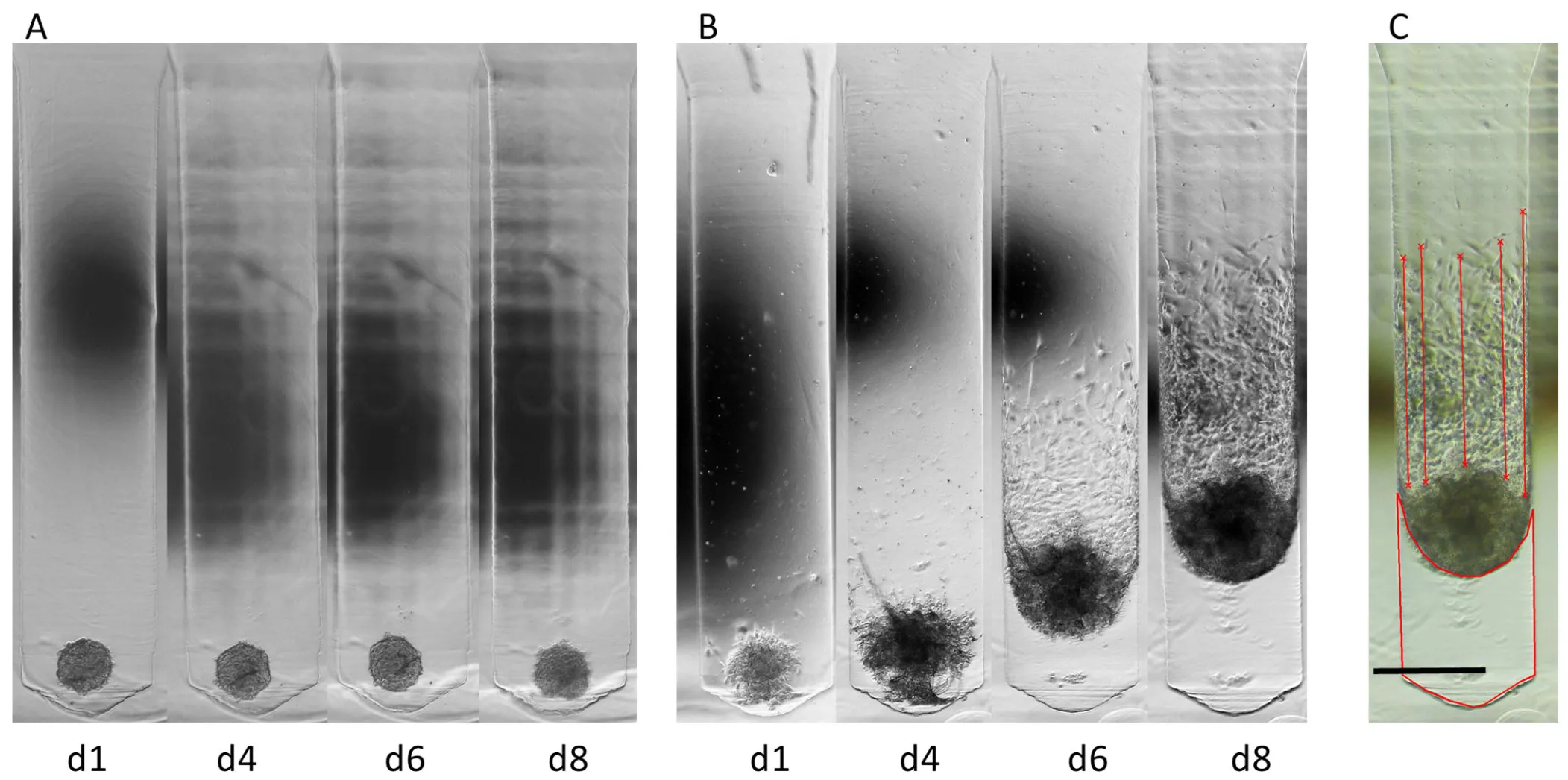
Bright-field time series of invasion from a cell aggregate, with the measurement overlaid.
Invasion over eight days (U-251 glioblastoma cells): without matrix the aggregate stays compact (left); with ECM the cells migrate into the matrix (middle). On the right, the readout: invasion distance (lines) and cleared matrix area (outline). Source: Thomsen et al., Methods Protoc. 2026, 9(4):112 · CC BY 4.0

What it measures

Three readouts, one compartment.

Migration, invasion and matrix degradation from one defined starting point, instead of three separate assays.

Invasion distance · µm

Invasion

Distance cells advance from the aggregate into the matrix.

Migration · µm

Single-cell migration

Movement of individual cells, separate from the aggregate.

Cleared area · µm²

Matrix degradation

Area the cells clear within the collagen or ECM.

How it works

From aggregate to measurement in four steps.

Bright-field microscopy is enough, no special equipment. Letters refer to the panels of the figure.
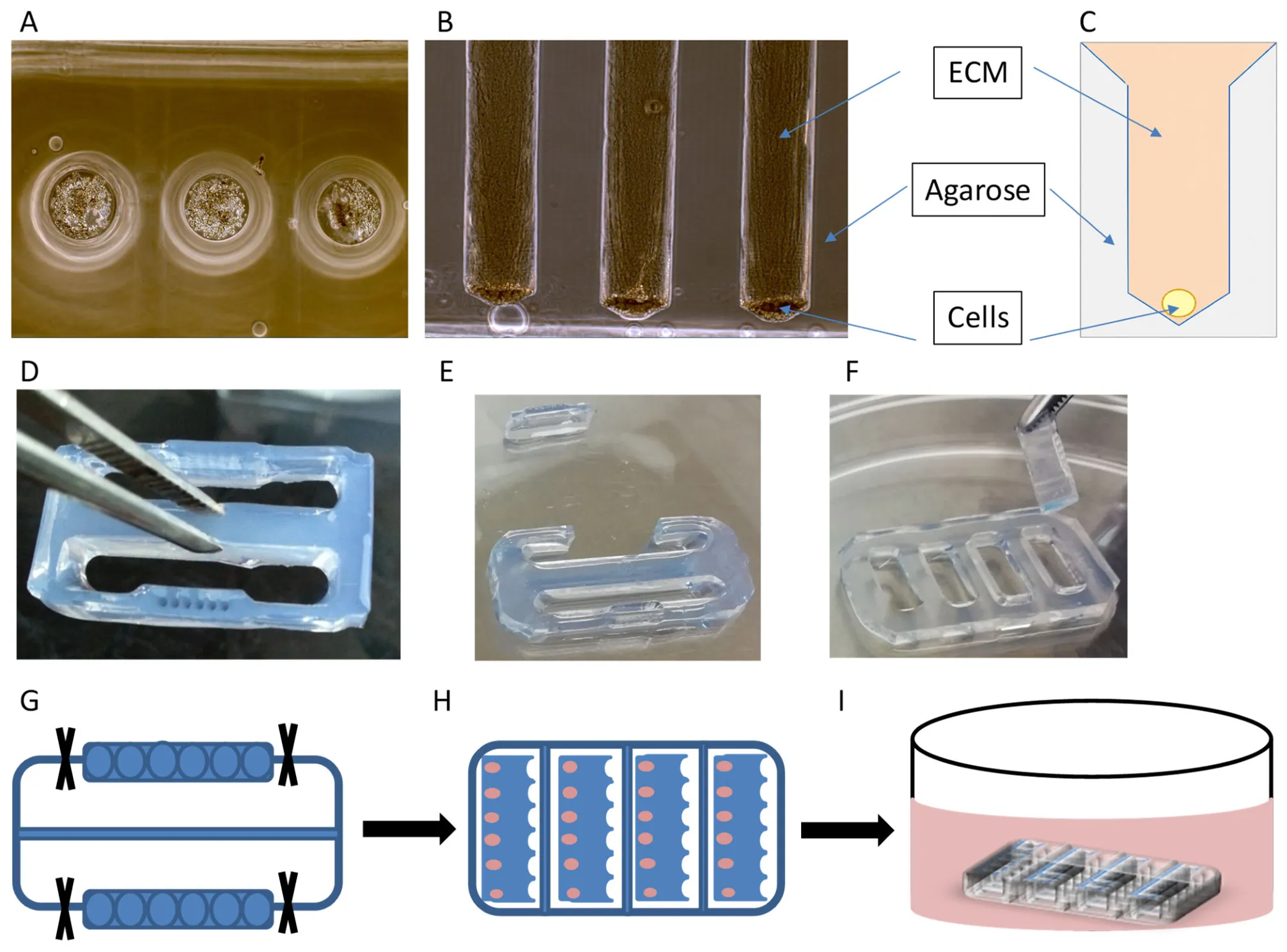
Multi-panel figure of the assay principle: cavities with cells and matrix plus schematic drawings.
Principle: cells form a compact aggregate at the base of each cavity, with collagen or ECM gel added on top (schematic, top right). For culture and imaging the strips sit in the parking pockets. Source: Thomsen et al., Methods Protoc. 2026, 9(4):112 · CC BY 4.0
  1. 01

    Seed the cells

    Seed tumor cells into the cavities of the gel carrier; they form a compact aggregate (fig. A).

  2. 02

    Add the matrix

    Add collagen type I or ECM gel; it polymerizes over the cells (fig. B, C).

  3. 03

    Transfer into the parking pockets

    Cut the loaded carrier into its two strips (the halves of the carrier), set them in the parking pockets, then culture and image in bright field over days to weeks (fig. D–I). Add irradiation or compounds here.

  4. 04

    Quantify

    Measure invasion distance, single-cell migration and cleared matrix area from the images.

Why this assay

Defined, reproducible, low-cost.

Between simple 2D/Transwell assays and complex organ-on-chip systems.

Defined start

Each cavity fixes the aggregate in the same position, unlike variable spheroids.

Three readouts at once

Migration, invasion and matrix degradation in one compartment.

No special equipment

Bright-field microscope only. No microfluidics, bioprinting or Matrigel drops.

Adherent and non-adherent

Also cell types that cannot cross a Transwell membrane.

For drugs and radiation

Shown with a protease inhibitor and clinical irradiation.

Flexible matrix

Collagen type I or ECM gel, depending on your question.

Validated

Invasive vs. weakly invasive, clearly resolved.

MDA-MB-231 invaded and degraded matrix far more than MCF7. A protease inhibitor and 2 Gy irradiation both reduced invasion.
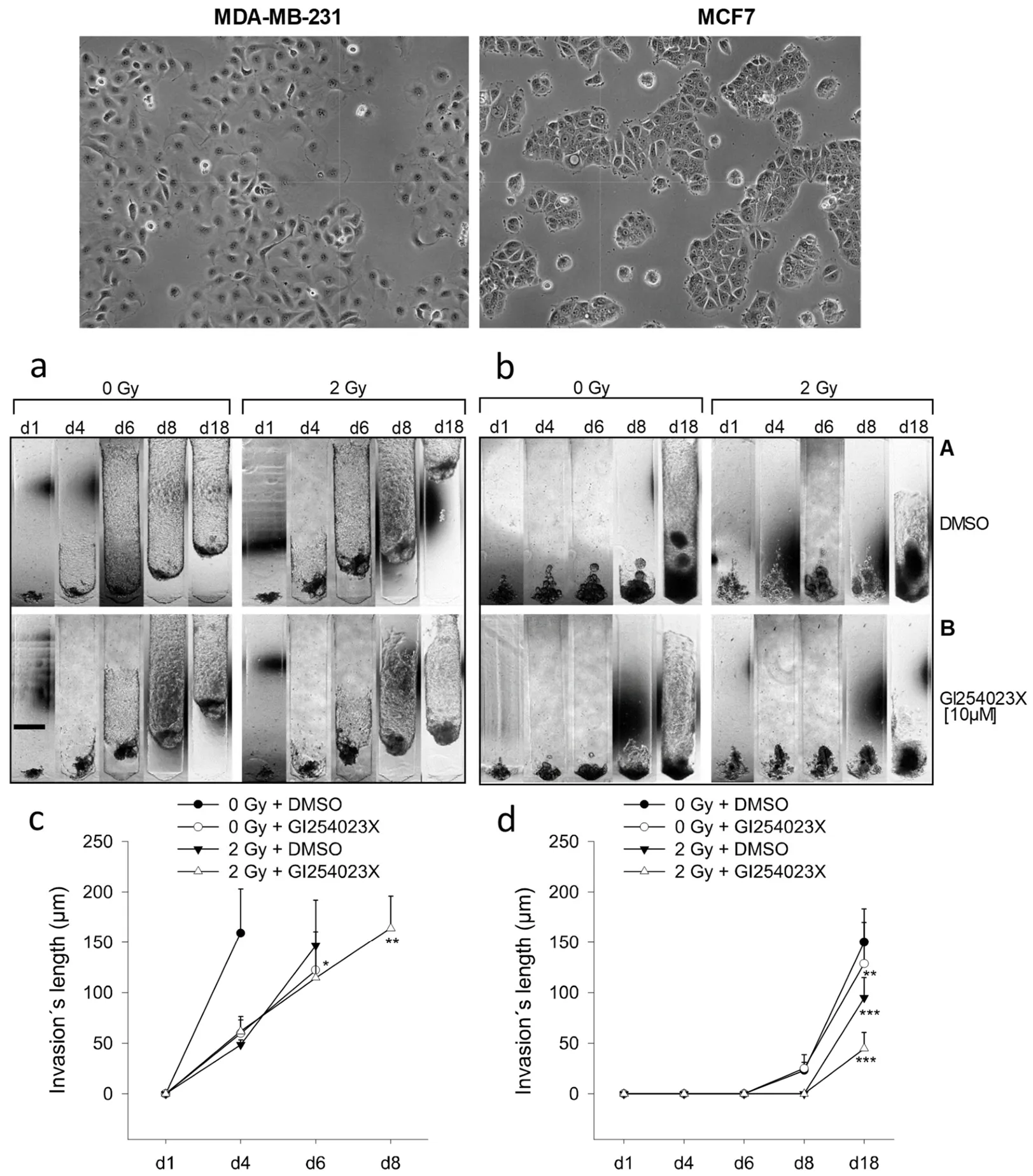
Comparison of two breast cancer cell lines with invasion images and quantification graphs.
Validation: the assay separates highly invasive MDA-MB-231 from weakly invasive MCF7 breast cancer cells. A protease inhibitor and 2 Gy irradiation reduce the measured invasion distance (graphs, bottom). Source: Thomsen et al., Methods Protoc. 2026, 9(4):112 · CC BY 4.0

Specifications

The carrier at a glance.

Delivered ready to use.

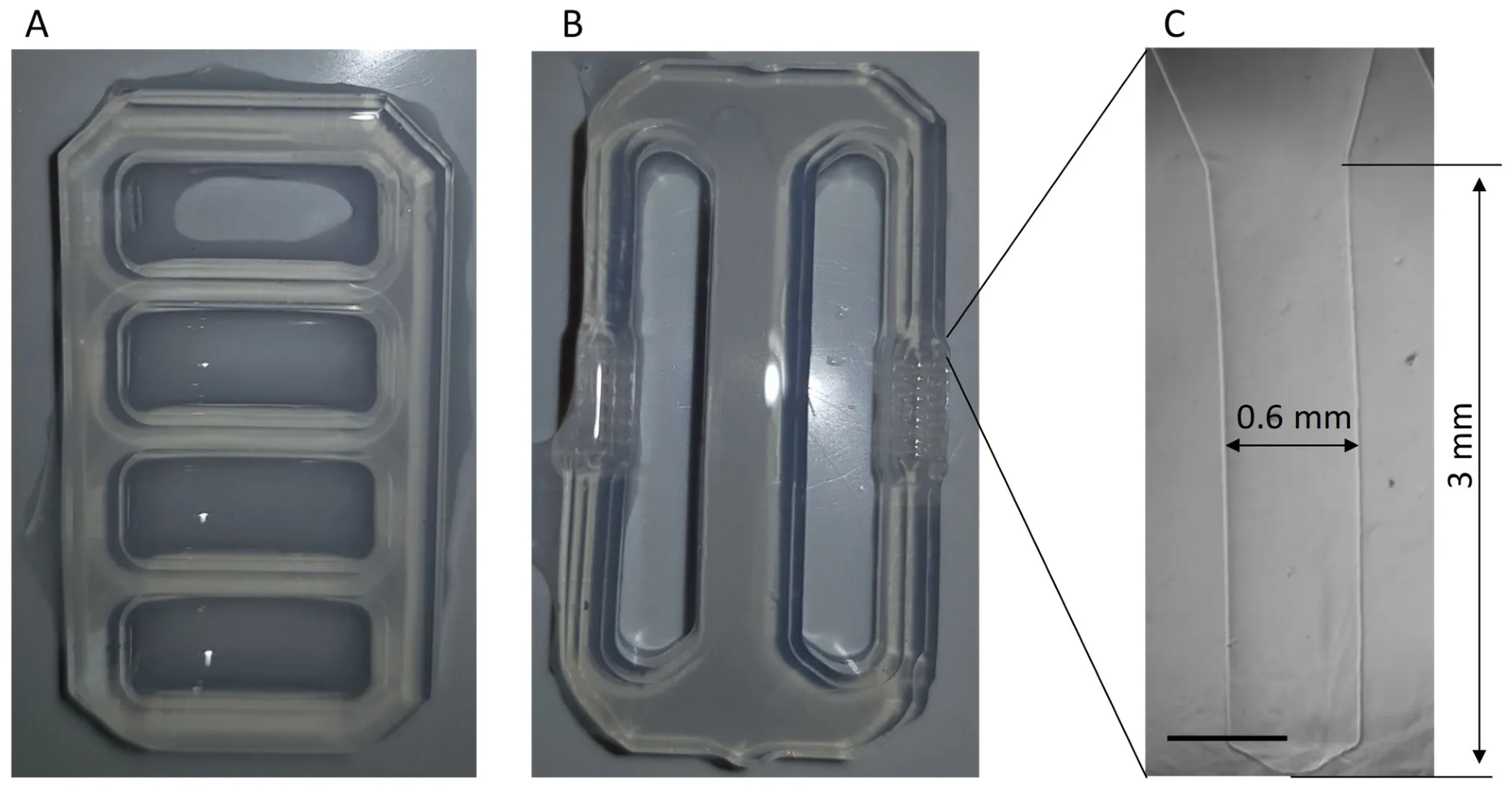
Photographs of the translucent agarose components of the Invasion Assay with a close-up of one cavity.
The ready-to-use agarose components: the gel carrier with six cavities per strip (centre) and the parking pocket that holds it (left). On the right, a single cavity in detail, about 0.6 mm wide and roughly 3 mm deep. Source: Thomsen et al., Methods Protoc. 2026, 9(4):112 · CC BY 4.0
Base material
2.8% agarose, animal-component-free
Cavities
0.6 mm diameter, about 3 mm deep, 6 per strip
Loading channel
Common channel, 15 µL
Components
Gel carrier and parking pocket, each about 28 × 12 mm
Readout
Bright-field microscopy, time series over days to weeks
Metrics
Invasion distance, single-cell migration, cleared matrix area
Matrix
Collagen type I or ECM gel (user-supplied)
Delivery
Ready to use, UV-decontaminated, stored in PBS

Peer-reviewed

Published in Methods and Protocols.

Full method, materials and validation data, open access. First author: Andreas R. Thomsen.

Open publication (DOI)

Research use

Bring the Invasion Assay into your lab.

Tell us your cell lines and question. We coordinate carriers and documentation with you.

Contact Thom-Med